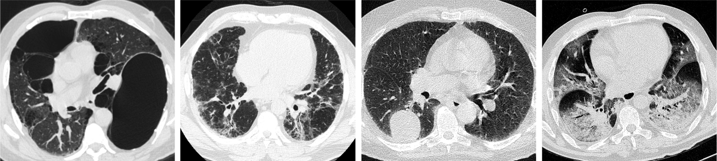
We specialize in using computed tomography (CT) imaging to produce high-resolution volumetric reconstructions of the lungs. We use CT imaging to understand normal and diseased lungs including chronic obstructive pulmonary disease (COPD), idiopathic pulmonary fibrosis, lung cancer, and COVID-19. Our lab develops advanced image analysis algorithms to improve automated quantification of image-based biomarkers and identification of pulmonary structures.
Lung Segmentation
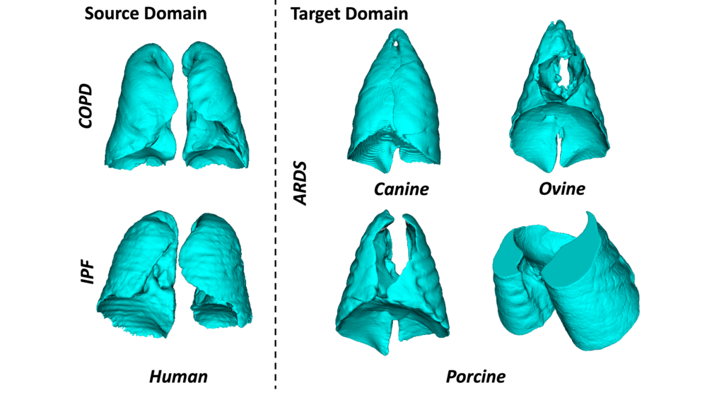
Lung segmentation is a necessary initial step for quantitative CT analysis in thoracic images. We have developed a robust method which is capable of accurately segmenting lungs across many pulmonary diseases including COPD, IPF, lung cancer, COVID-19, and acute lung injury. Furthermore, our method is capable of segmenting lungs of multiple species despite large variation in global lung shape and appearance.
Fissure Detection
The pulmonary fissures are the boundaries that separate the lungs into lobes. There are three main fissures that separate the the lungs into five lobes.
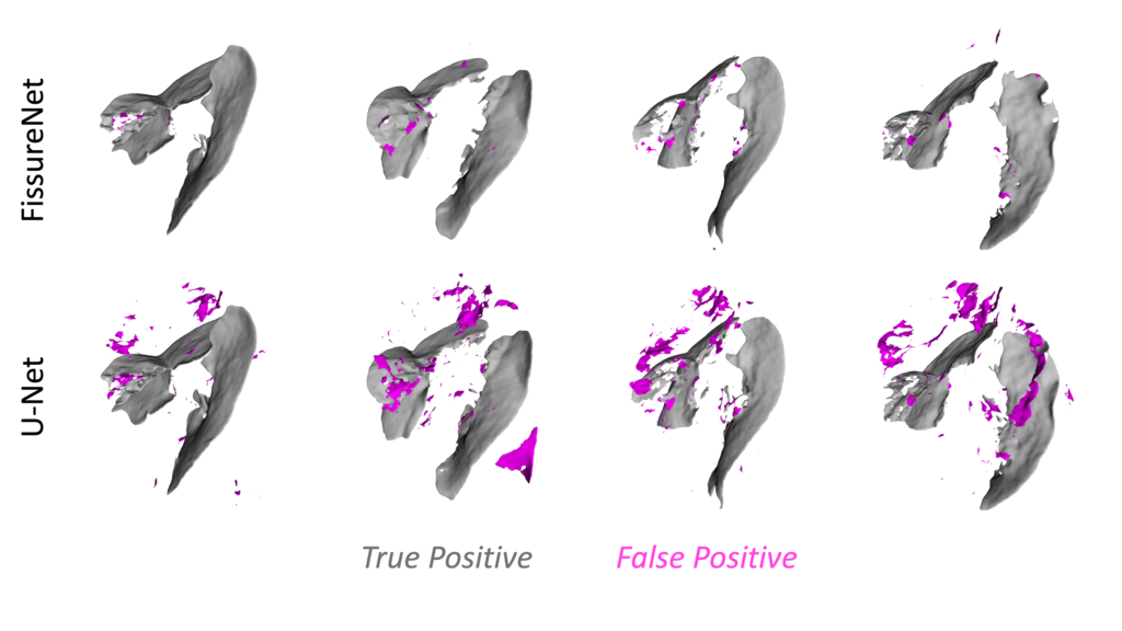
Detecting fissures in CT images is a challenging task as the fissure appears as a very thin surface-like structure, fissures can be incomplete or missing, and the appearance of fissures can be altered from disease.
We proposed a deep learning approach called FissureNet which is capable of detecting weak fissures and achieves a low false positive rate compared to other methods.
Lobe Segmentation
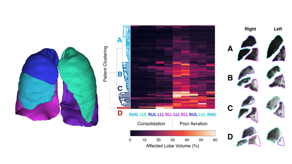
The lungs are divided into five compartments called lobes, which are functionally independent given complete fissures. Lobe segmentation is an important step for regional analysis in lung diseases.
We have used lobe segmentation combined with hierarchical clustering to identify four subgroups of COVID-19 based on lobar quantification of consolidation and poor-aeration.

Vessel Segmentation and Sizing
The pulmonary vessels form a complex branching structure to transport blood into and out of lungs lungs. Alterations in the pulmonary vasculature lead to changes in perfusion and thus lung function.
We developed an automated method to segment the total pulmonary vasculature and quantify the size of the vessels. We found a significant difference in the ratio of small vessels to large vessels in COPD rapid progressors compared to non progressors.
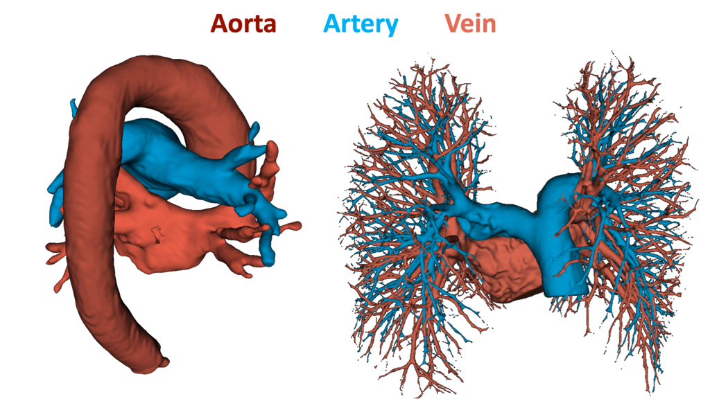
Aorta-Artery-Vein Segmentation
The pulmonary vessel tree can be divided into the arterial tree (pre-capillaries) and venous tree (post-capillaries). The arterial tree carries unoxygenated blood to the heart to the lungs and the venous tree carries oxygenated blood from the lungs to the heart.
Segmentation of the pulmonary arteries and veins enables more specific biomarkers to be identified to characterize pulmonary diseases.
We are utilizing contrast-enhanced dual-energy CT scans with deep learning to automatically segment the venous and arterial trees in the lungs and mediastinum in both contrast and non-contrast scans.