The primary function of the lungs is gas exchange, oxygen is brought into the lungs from the environment and waste gases are removed. This process requires matching of air flow and blood flow in the lungs, or ventilation and perfusion. We specialize in using advanced imaging modalities to image regional ventilation and perfusion and in developing image processing tools to analyze these images. The unique combination of our structural and functional imaging tools allows us to characterize structure-function relationships in normal and diseased lungs.
Ventilation: Image Registration
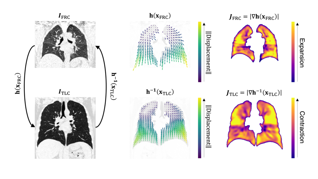
Image registration is the process of finding a transformation that aligns multiple images to a common reference space. Image registration between images at different lung inflation levels allows us to observe change in aeration at a specific voxel.
The displacement field can also be analyzed to quantify lung mechanics such as local tissue volume change or Jacobian. We use image registration and Jacobian images as a surrogate for regional ventilation, or air flow, in the lungs.
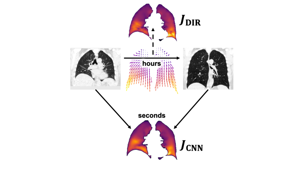
Ventilation: CT-to-Jacobian Translation
Image registration is an iterative process that can take several hours and can fail to converge to the global minima. We developed a deep learning framework that directly predicts the Jacobian image from a pair of CT images. This allows the Jacobian image to be produced in seconds.
This method was extended to predicting the Jacobian image from a single CT image, enabling its use for cases where only a single CT image is available. This is a task that is impossible in the image registration framework, yet is within reach for deep generative models.
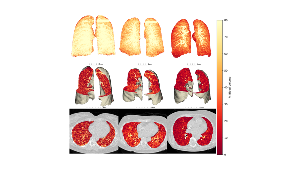
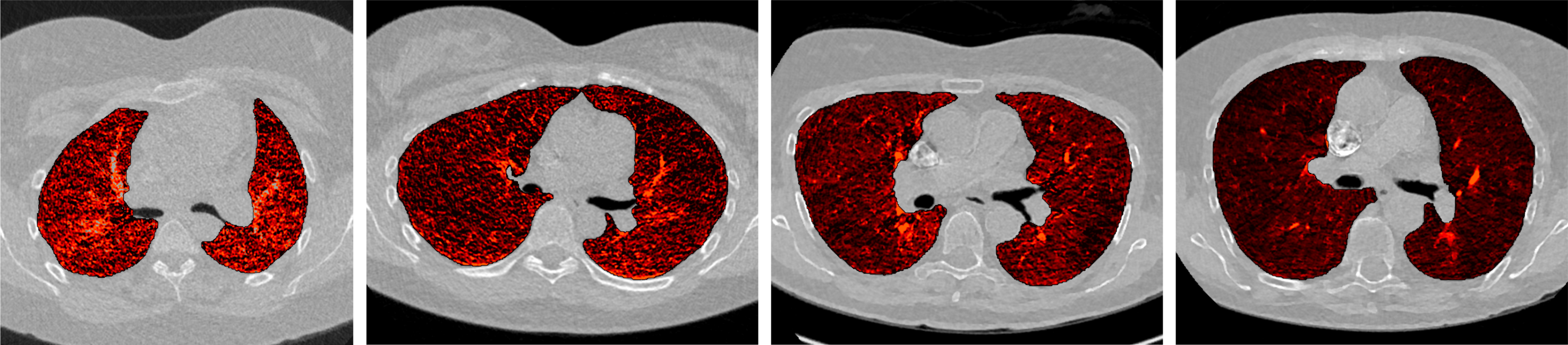
Perfusion: Dual-Energy CT
Dual-energy CT uses two distinct X-ray energy spectra to simultaneously acquire a low-energy CT image and a high-energy CT image. This enables the extraction of materials that have different X-ray attenuation properties at the two energies.
Dual-energy CT combined with iodine blood contrast allows the assessment of perfused blood volume in the lungs, a surrogate for pulmonary perfusion. We utilize dual-energy CT with contrast to quantitatively characterize and spatially resolve dysregulated pulmonary perfusion in various lung conditions including COPD and COVID-19.